Deformable models
Dynamic Deformable Models for 3D MRI
Heart Segmentation
Collaboration : Dr. John Wood, LA Children's Hospital ; Dr. Igor Guskov University of Michigan; Dr. David Breen, Caltech
Undergraduate student: Josh Bao, Caltech
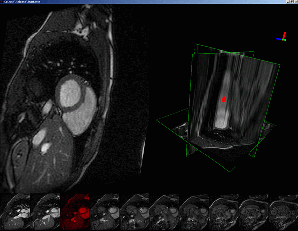
Cine magnetic resonance imaging has become the gold-standard for measurement of myocardial mass and function. Typically, cross-section images at 10-12 anatomic levels are imaged at 16-32 equally spaced points throughout the cardiac cycle. Current state-of-the-art commercial tools use semi-automated boundary detection to trace endocardial and epicardial boundaries at their maximum (end-diastolic) and minimum (end-systolic) frames. The traced contours are then combined to produce boundary representation of the volumes. Typical user time for such processing varies between 20-60 minutes for right and left ventricular volumes and ejection fractions. Since the post-processing is so labor intensive, all data except the end-diastolic and end-systolic frames are ignored, discarding important function information regarding ejection and filling rates. These dynamic variables offer additional insight into myocardial systolic and diastolic performance.
 |
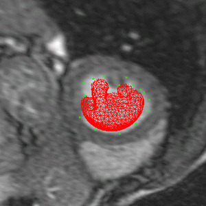 |
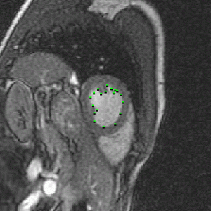
We have developed software tools to automatically track myocardial boundaries through all the time steps. In our system the surface model is initialized with a user assistance by specifying control points at end-systolic and end-diastolic frames. The time evolution of the tracked surface is then computed using deformable model framework. A deformable model is an elastic body that deforms under the applied forces until it fits to the data. These forces consist of image forces that attract the model to the image features and elastic forces due to deformation energy of the model that ensure its smoothness. Additionally, deformable model use a priori knowledge about the location, size and shape of the ventricles,and allow an easy interactive mechanism for the user to constrain and navigate the development of the model.
 |
 |
 |
More details on this project can be found in the paper:
L Zhukov, Zh. Bao, I. Guskov, J. Wood and D. Breen
Dynamic Deformable Models for 3D MRI Heart Segmentation
SPIE Medical Imaging 2002 , February 2002.